
Found a New Pet Lump? Record It for Your UK Vet
Quick answer: Book veterinary advice for every new lump, even if it looks harmless or resembles an older one. Note its exact location, photograph it with a size reference, measure length and width gently, and record pain, heat, colour, texture, bleeding, or discharge. Do not squeeze, pierce, apply human creams, or delay the appointment while waiting to see what happens.
Lumps and swellings have many causes, and different conditions can look similar from the outside. A useful home record helps the vet see change over time, but only veterinary assessment—and sometimes a sample—can identify what a lump is.
1. Treat Every New Lump as New Information
Do not assume a new lump is a cyst, fatty lump, bite, abscess, or tumour based on touch or an internet image. PDSA advises having all new lumps examined by a vet. A pet that has had a harmless lump before can still develop a different condition elsewhere.
Book advice when you first find it and tell the practice how long it may have been present. If you are unsure whether a bump is normal anatomy—such as a nipple, joint, rib, or microchip—do not repeatedly manipulate it. Photograph the location and ask.

2. Spot Reasons for Faster Veterinary Help
Tell the practice promptly if the lump grows quickly, changes shape or colour, becomes hot or painful, bleeds, opens, smells, or leaks fluid. Faster assessment is also sensible when the pet is unwell, feverish, unusually quiet, not eating, losing weight, breathing differently, or persistently licking or scratching the area.
Seek urgent veterinary help for breathing difficulty, collapse, severe bleeding, rapidly expanding facial or throat swelling, a suspected serious allergic reaction, or another life-threatening change. Do not wait for a routine appointment in an emergency.
Greenmount Vets Portdown
PortadownArmagh City, Banbridge and CraigavonNorthern Ireland
72 Gilford Rd, Portadown, Craigavon BT63 5EG, UK
3. Map the Exact Location
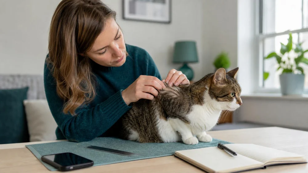
Use a simple body outline or written landmarks: “left side of chest, two finger-widths behind the front leg” is more useful than “on the body.” Note whether the lump is in the skin, just beneath it, attached deeper, or impossible to distinguish without causing discomfort.
If there are several lumps, assign each one a number and record it on the body map. Do not rely on memory or use the same photo label for different areas. The map helps the vet match each measurement to the correct lump.
4. Take Comparable Photographs
Use good natural light and gently part fur without pulling. Take one wider image showing location and one close view. Place a ruler beside—not pressed into—the lump when safe. Keep the camera at a similar angle and distance for later comparison.
A photo cannot show depth or diagnose tissue, and perspective can exaggerate change. Save the date and body-map number with each image. If the pet resists, becomes distressed, or may bite or scratch, stop and let the veterinary team examine it.
5. Measure Without Squeezing
Measure the longest visible length and the width at right angles in millimetres or centimetres. If the lump is raised and your vet asks, record approximate height without pinching. Use the same unit every time.
Do not repeatedly press to decide whether it is “hard,” “fixed,” or “fluid-filled.” Gentle observation once is enough. Excess handling can cause pain, inflammation, bleeding, or misleading change—especially if the area is infected or fragile.
6. Describe What You Observe
Record factual features rather than a home diagnosis:
- date first noticed and whether onset seemed sudden or gradual;
- colour and whether skin or fur over it looks different;
- smooth, uneven, soft, firm, or impossible to assess gently;
- warmth, tenderness, itch, licking, or effect on movement;
- bleeding, crust, opening, odour, or discharge;
- changes in appetite, energy, weight, toileting, or breathing;
- recent injection, bite, wound, tick, surgery, or known injury nearby.
Tell the vet about medicines and existing diagnoses. Do not stop prescribed treatment because a lump appears unless the prescriber advises it.
7. Avoid Home Treatment and Damage
Do not squeeze, lance, drain, cut, tie off, burn, or freeze a lump. Do not apply human wart removers, steroid creams, essential oils, antiseptics, or leftover animal medicines without veterinary direction. Some products are toxic when pets lick them, and treatment can alter the surface before examination.
Prevent licking or rubbing using only a method the practice recommends. Keep a bleeding or open area clean and protected as directed, and ask the vet whether it needs same-day care. Never use a tight bandage over an unidentified lump.
8. Prepare for the Appointment
Bring the body map, dates, measurements, photos, symptom notes, medication list, and relevant history. Tell the practice if the pet is fearful, reactive, or painful so handling can be planned. Follow instructions about feeding before the visit; do not fast a pet unless the practice tells you to.
Ask what the examination can and cannot determine. Because many lumps look similar, the vet may recommend a fine-needle sample, biopsy, imaging, removal, laboratory testing, or monitoring. Ask what each option is intended to answer, its risks, cost, timing, and what result would change the plan.
9. Monitor Only With Veterinary Guidance
Home monitoring is appropriate when the vet has examined the lump and given a plan. PDSA suggests checking an existing lump at least weekly when monitoring has been recommended, using photos, length and width, and notes on shape, texture, colour, pain, itch, or discharge.
Confirm the review interval and triggers for an earlier appointment. Contact the vet if the lump grows rapidly, changes shape, colour, or texture, starts bleeding or discharging, or becomes painful. “Watch it” should have a date and clear escalation rules—not an indefinite delay.
10. Review Common Lump Questions
Can a vet identify a lump by looking at a photo?
A photo helps triage and track change but often cannot identify the tissue. PDSA notes that lumps can look similar externally and may require sampling for diagnosis.
Is a soft, movable lump harmless?
Texture and movement alone cannot prove that. Arrange veterinary assessment rather than labelling it at home.
Should I wait to see whether it grows?
Contact the vet when you find a new lump. Monitoring should begin after the practice has advised that approach, not instead of initial assessment.
What if my pet has many lumps?
Map and number each one and tell the vet which is new or changing. Each new lump can be different, so do not assume one previous test covers all future lumps.
Owner checklist: book veterinary advice; map the site; take overview and close photos; measure length and width gently; note surface and whole-pet changes; avoid squeezing or home remedies; bring a clear record; and agree on review and escalation rules.
Evidence notes: This guide follows PDSA’s UK lump-monitoring advice, which says new lumps should be examined and describes weekly checks for lumps a vet has asked an owner to monitor. Its cat lump guidance explains why external appearance may not be enough and why sampling may be needed.
This article is general pet-care information, not a diagnosis. Contact a veterinary practice for every new or changing lump and whenever your pet seems unwell.
 Spen Corner Veterinary Centre4.0 (128 reviews)
 Pets Corner4.0 (352 reviews)
 Barn View Cattery5.0 (69 reviews)
 Melton Vets4.0 (260 reviews)
 Calder Vets in Mirfield4.0 (43 reviews)
 Abbey House Vets in Halton4.0 (108 reviews)
 UK Pet Owner’s Guide to Adoption for Reptiles
 Top 10 Weight Management Tips for Exotic Pets Owners in the UK
 Best Practices for Puppy and Kitten Care in Cats: A Complete Guide
 Behaviour Training Tips for Puppies: What UK Pet Owners Need to Know
 How to Handle Emergency Situations with Your Pet | Pet Safety Tips
 Why Is My Cat Hiding Under the Bed All Day? Fear or Illness Explained